Address
1. 70 Asklipiou Street, Mytilene, 81100
2. 7 Myraktis Street, Nea Smyrni, 17121
Working Hours
Mon-Fri: 09:00-21:00
Saturday:10:00-14:00

Lorem ipsum dolor sit amet, consectetur adipiscing elit. Nam bibendum ultrices arcu ac vestibulum. Nam nec tempus quam.

[fusion_separator style_type=”none” sep_color=”” border_size=”0″ icon=”” icon_circle=”” icon_circle_color=”” alignment=”center” hide_on_mobile=”small-visibility,medium-visibility,large-visibility” class=”” id=”” top_margin=”10px” bottom_margin=”10px” /]

[fusion_button link=”https://physiogalinos.gr/our-history/” text_transform=”” title=”” target=”_self” link_attributes=”” alignment=”left” modal=”” color=”default” button_gradient_top_color=”” button_gradient_bottom_color=”” button_gradient_top_color_hover=”” button_gradient_bottom_color_hover=”” accent_color=”” accent_hover_color=”” type=”” bevel_color=”” border_width=”” size=”” stretch=”default” shape=”” icon=”” icon_position=”left” icon_divider=”no” animation_type=”” animation_direction=”left” animation_speed=”0.3″ animation_offset=”” hide_on_mobile=”small-visibility,medium-visibility,large-visibility” class=”” id=””]Our History[/fusion_button]

διάστρεμμα ποδοκνημικής

Chronic Lateral Ankle Pain

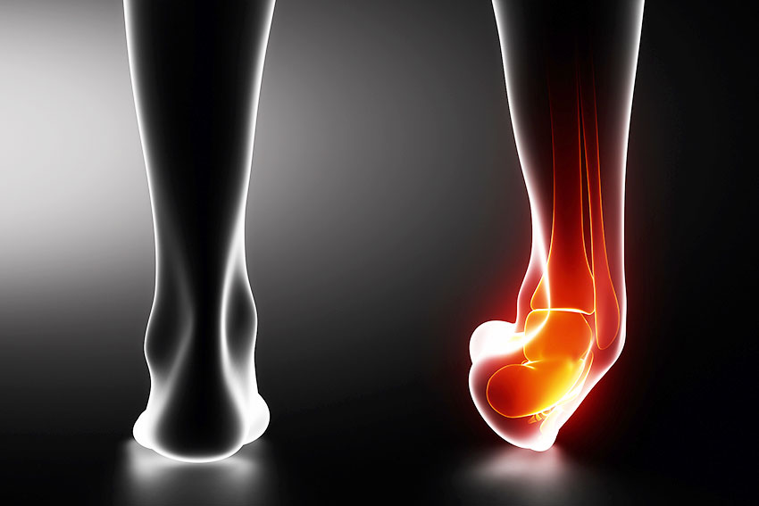
The ankle is the joint that forms between the tibia and the ankle. This joint is located in the lower part of the lower limb and connects the tibia to the foot.

The bones are connected to each other and fixed with connective tissue strips, which are called ligaments. The ligaments guide the movements of the joints and do not allow the bones to move too far apart. The joints are normally very elastic and when stretched they return to their normal length without any other problems. However, when due to a violent movement the bones are significantly separated from each other then the ligaments are significantly stretched and it is possible to cut. In this case the patient feels intense discomfort.

In the ankle joint there are ligaments on the outside and on the inside. On the outside of the ankle there are 3 tapered ligaments and on the inside there is a flattened ligament, the medial lateral ligament. The 3 ligaments on the outside of the ankle have different names depending on the area of ​​protrusion and bulge. The more severe the sprain, the more joints have been injured.

Recurrent or permanent (chronic) pain in the outside of the ankle often develops after an injury such as a sprained ankle. However, many other conditions can also cause chronic ankle pain.

Symptoms

The ankle shows swelling (swelling) almost immediately after turning the foot. The swelling is usually more severe on the outside of the ankle and may be accompanied by hematoma. At the same time charging the limb is painful or even impossible.

The symptoms concern:

  • Pain usually on the outside of the foot (the pain may be so severe that the patient has difficulty walking, in some cases the pain is a permanent, hazy pain)
  • Difficulty walking on uneven ground or toes
  • Feeling of instability
  • Swelling
  • Stiffness
  • Sensitivity
  • Repetitive ankle sprains

Causes

The most common cause of a persistently sore foot is inadequate healing after an ankle sprain. When you get a sprained ankle, the ligaments between the bones stretch. Without a complete recovery, the ligaments and surrounding muscles may remain weak, leading to recurrent instability. As a result, you may experience further ankle injuries.

Other causes of chronic pain include:

  • Injury to the nerves that penetrate the ankle. The nerves can be stretched or injured by a direct hit or by constant pressure while the foot is trapped.
  • Cut or inflamed tendon.
  • Ankle arthritis.
  • Fracture of one of the bones that make up the ankle joint.
  • Inflammation of the synovial membrane.
  • Development of scar tissue in the ankle after a sprain. The scar tissue takes up space in the joint, squeezing the ligaments.

Diagnosis

Ankle x-rays are usually requested by a specialist. An x-ray of the other leg may also be needed so that the doctor can compare the injured to the non-injured ankle. In some cases, additional tests may be needed, such as a bone scan, CT scan, or MRI scan.

Treatment

The treatment will depend on the final diagnosis and should be individualized to the needs of the individual. Both conservative and surgical methods are used.

The conservative method includes:

  • Anti-inflammatory drugs such as aspirin or ibuprofen to reduce swelling.
  • Physiotherapy, which includes natural means (Diagnostic pump, TECAR, currents, LASER etc) & exercises (on a balance disc, with the aim of strengthening the muscles, restoring range movement, and increase the perception of the position of the joint.)
  • Ankle splint or other support.
  • Injection of a steroid medication.
  • In the event of a fracture, immobilize to allow the bone to heal.

Sources
Harmon K.G., (2007). Which support is best for first-time ankle sprains? Clin J Sport Med, 17 (4): 333 – 334.
Weber J.M., & Maleski R.M., Conservative treatment of acute lateral ankle sprains, Clin Podiatr Med Surg, 19 (2): 309 – 318.
Φυσικοθεραπεία Πάρκινσον

Physiotherapy for Parkinson’s Patients

Parkinson’s disease is one of the most common neurological diseases. As in all degenerative conditions, the onset is insidious and once the patient seeks medical advice, it is often possible to report a history of a few months or even years. This disease can be characterized as a disease of wear and tear. That is, it occurs when certain brain cells can no longer produce dopamine. Because of this, the chances of developing Parkinson’s increase with age.

Possible causes of the disease are atherosclerosis, alcoholism, drug use, metabolic disorders, toxic substances (such as manganese and MPTP toxin), medication (mainly neuroleptics), brain tumors, and and premature aging of the neurons of the substantia nigra. Finally, the existence of an inherited predisposition to the disease has not been proven and does not seem possible.

So far there is no cure for the disease or a way to prevent the disease from progressing. But there are several drugs that significantly improve the symptoms for many years, thus improving the quality of life. Drug therapy is based on replenishing dopamine levels in the brain. There is also surgery, when the drugs work but do not constantly control the symptoms. Patients must meet specific criteria to be eligible for surgery. Exercise has also been shown to help a lot in controlling symptoms and well-being.

Symptoms

The disease first appears with the combination of two symptoms, bradykinesia and stiffness or tremor and stiffness, and as it progresses, all the characteristic manifestations of the disease appear slowly. It most often occurs in the sixth and seventh decade of life and affects both sexes equally. It can, however, occur at almost any age although it is very rare under thirty. As age increases, so does the frequency.

Essentially, the onset of the disease is slow and the course progressive. The initial symptoms are not typical. Diffuse pains are observed, mainly in the shoulders, due to the stimulation of the joints, feeling of fatigue and reduction of daily activities. The patient complains that he has become sluggish and cumbersome, but often attributes the discomfort to old age. When the disease is established, then the characteristic four symptoms are observed: bradykinesia, tremor at rest, stiffness, as well as loss of corrective reactions.

Non-motor symptoms, such as constipation, urinary disorders (frequent urination, nocturia, urination), olfactory disorders and depression, are also common, which may precede motor manifestations. Over time, and especially when the disease begins in old age, dementia (about half of patients after 15 years of follow-up) is very common, often accompanied by visual hallucinations.

Diagnostic Approach

The diagnosis of the disease is made only by clinical examination. An experienced neurologist is able to diagnose the disease through the patient’s symptoms and clinical examination. There is no laboratory test to confirm the disease.

There are only tests recommended by a neurologist to rule out other conditions similar to Parkinson’s disease.

Parkinson’s disease is divided into idiopathic and secondary.

Parkinson’s idiopathic disease is a progressively developing disease with the main manifestations being restlessness, stiffness, sluggishness and loss of reflex postures (corrective postures). There must be at least two of the above main symptoms in practice to diagnose the disease.

Secondary Parkinsonism presents a similar clinical picture to the idiopathic and is caused by factors such as infections (viral encephalitis), toxic substances, brain tumors, etc.

Every patient has a different development. Still, the evolution can stop for some other time. It can be slow and mild, or for no apparent reason lead to significant deterioration.

Prevention

To date, no reliable method of preventing the disease has been found. Therefore, early or even early diagnosis does not help anything. There is no way to reverse or even delay the progression of the disease. Neither early medication nor any exercise or special diet has been shown to slow the progression of long-term disability caused by the disease.

On the contrary, there is evidence that early administration of dopamine or similar dopamine-mediated drugs, called dopamine agonists, may damage specific cells. For this reason there is no need to rush into the treatment of the disease.

Self-restraint and the most conservative medication possible are the best strategy in Parkinson’s disease.

Physiotherapy Treatment

The current model of physiotherapy intervention in patients with Parkinson’s is based on the assumption that normal movement can be acquired through teaching. Knowledge of the features of motor disorders in Parkinson’s patients is the starting point for designing a rehabilitation program. Most patients with Parkinson’s have difficulty walking at some stage of the disease. The use of external stimuli and cognitive strategies are the main therapeutic options of the physiotherapist for the gait disorder. Consequently, the elimination of falls is an important goal of physiotherapy, especially in patients in the later stages of the disease. Also, the prevention of muscle weakness and atrophy, limited range of motion and reduced ability to exercise, is a major goal of physical therapy in the Parkinson’s patient. Continuous physiotherapy is not necessary, but frequent meetings and advice are valuable.

Aging, any concomitant pathological conditions and secondary adaptive changes in the musculoskeletal and cardiovascular systems are also very important issues in the design of the physiotherapy program. Because Parkinson’s generally progresses slowly, patients and their families need to be supported in developing programs that should be implemented during long-term treatment. This support can help them take on more responsibility for their health and well-being in general.

Sources
by Goede G.J.T., Keus S., Kwakkel G., & Wagenaar R., (2001). The effects of physical therapy in Parkinson’s Disease: A research synthesis, Archives of Physical Medicine and Rehabilitation, 82 (4): 509 – 515.
Kwakkel G., de Goede G.J.T., & van Wegen E., (2007). Impact of physical therapy for Parkinson’s disease: A critical review of the literature, Parkinsonism & Related Disorders, 13 (3): 478 – 487.

κήλη μεσοσπονδύλιου

Herniated Disc

Spinal disc herniation (herniated disc) is a common condition mainly in the lumbar spine but also the other degrees of the spine. At the same time it is and is the most common cause of pain in both the upper and lower extremities.

The intervertebral discs are located between the vertebrae and absorb the vibrations of the spine. Each disc is hard on the outside (fibrous ring) and contains a soft core inside, called the gel nucleus. Herniated disc occurs when the hard ring ruptures and the nucleus from the inside of the disc slides into the spinal canal and presses on a nerve.

A hernia can occur in any part of the spine, but the highest frequency is clearly seen in the lumbar spine, then in the cervical spine and much less frequently in the thoracic spine.

The risk factors for herniated disc are:

  • Age: As we get older, our discs become damaged (corresponding to the deterioration of the skin and the appearance of wrinkles).
  • Lifestyle: Lack of exercise, being overweight and smoking lead to poor intervertebral disc function.
  • Poor posture: Poor posture and repeated weight lifting or waist turns cause stress on the discs.

Symptoms

This disease (disc disease) often occurs in young people, but also in middle and old age. Symptoms may include lower back pain (back pain) and leg pain (sciatica), and sometimes weakness (paralysis) and numbness of the leg.

The symptoms that appear vary depending on the severity of the hernia and its location.

Patients complain of acute or chronic low back pain (back pain), while in nerve root involvement there may be reported pain, numbness or paraesthesia in the lower extremities (sciatica).

Also often accompanied by muscle spasm, loss of lordosis in the waist and worsening of symptoms with coughing or sneezing. Sometimes analgesic scoliosis is created, ie distortion of the body, in the involuntary reaction of the person so that it does not hurt at the point where the root is pressed.

It should be mentioned here that in very serious situations, such as hippocampal syndrome, where there is weakness of the lower extremities, saddle type anesthesia, problems with urination, etc., immediate surgical treatment is required.

Diagnosis

Diagnosis begins with a history and clinical examination of the lower back (lower back) and lower limbs. The diagnosis is then confirmed by magnetic resonance imaging.

The diagnosis of hernia is made accurately by magnetic resonance imaging (MRI), but plain X-ray is also useful in differentiating from other conditions, such as tumors, and stenosis of the intervertebral space on X-ray is an indication of possible hernia.

However, this does not mean that anyone with a hernia should necessarily be in pain or undergoing treatment, as a large percentage remain asymptomatic or the pain disappears over time.

Dealing

Initial treatment of herniated disc should be conservative and focused on relieving pain and other symptoms. Research has shown that the vast majority of back pain is due to discogenic problems, with prominent projection or herniation of the intervertebral disc.

Drug analgesia, bed rest and conventional physical therapy using machines such as TENS, laser, diathermy and massage aim to reduce pain, but only provide a temporary relief and not a substantial treatment of the problem.
An appropriate program of exercises to strengthen the stabilizers and supporting muscles of the waist, seems to offer something more than the above means.

Physiotherapy Treatment

The McKenzie Method – Mechanical Diagnosis and Treatment (MDT) is a comprehensive system for the proper evaluation and treatment of back pain. An important advantage of the method, compared to the other approaches, is the prognosis from the first sessions, ie which incident will respond positively to treatment and which will not. This gives confidence and security to the sufferer and reduces the unnecessary waste of time and money on dubious treatments.

mckenzie method

The other great advantage of the McKenzie method is that it is based on Self-Healing, through special individualized exercises that are designed based on the appropriate assessment that precedes, while no machines or drugs are used. Thus, little by little, the patient becomes completely independent of the therapist, as he is trained for a correct attitude in everyday life and eventually learns to manage his condition on his own, preventing any relapses in the future.

The McKenzie method has avoided a very large percentage of planned surgeries for herniated disc and now the method has gained the recognition of orthopedic surgeons and neurosurgeons. Thus, surgery is chosen only when a proper conservative treatment fails to reduce the patient’s symptoms or when there are severe neurological lesions with pressure on the nerves or spinal cord.

Sources
Kontzaelias DA, (2011). Physiotherapy in diseases of the musculoskeletal system, Publications: University Studio Press, Thessaloniki.

ρήξη πρόσθιου χιαστού 1

Anterior Cruciate Ligament Rupture

The anterior cruciate ligament (ACL) is the main stabilizer of the knee joint. Its role is to protect the joint from the anterior displacement of the tibia in relation to the thigh, to control the rotation of the joint and its lateral displacements (stiffness, flexibility).

Its function provides significant stability to the joint during daily activities, running, changing directions and landing by jumping. Rupture of the anterior cruciate ligament involves injury to the ligament in the knee that connects the lower thigh to the upper tibia. It is characteristic that a patient with a rupture of the anterior cruciate ligament complains of instability of his joint but also of episodes where his knee slides anteriorly(παρεμβολη εικονας : anterior cruciate ligament rupture).

acl tear

Epidemiology
The incidence of anterior cruciate ligament injury in the knee is two to eight times higher in women than in men. In basketball in particular, women are four times more likely to be injured than men. This difference between the sexes is even greater in people attending military academies. Also, people who have had joint ligament repair have a higher risk of rupture in both the (most often) and healthy limb.

The incidence of anterior cruciate ligament injury is high in basketball, hockey, soccer, American football, gymnastics, skiing, and volleyball. Anterior cruciate ligament ruptures are constantly increasing due to the large participation of the population in these sports. Especially in skiing, anterior cruciate ligament injuries cover 25% to 30% of all knee injuries. Professionalism and the high level of sports today reinforce the above phenomenon.

Diagnosis
A good history combined with special tests can make the diagnosis without the need for additional tests. Measuring the relaxation of the ligament with special instruments enhances the clinical examination and provides an objective reference point for future comparisons. The most commonly used articulator is the KT-1000 which uses constant forces to determine the anterior tibial displacement.

ρήξη πρόσθιου χιαστού 2

Of great importance are the simple radiographs with which fractures of the knee must be ruled out (but also detached fractures in the protrusion of the ligament). Although usually not necessary for diagnosis, magnetic resonance imaging (MRI) is useful as it is more than 95% accurate in diagnosing anterior cruciate ligament rupture. It is also extremely useful in the search for concomitant lesions. Another imaging examination is the knee arthroscopy which, in addition to being diagnostic, also has therapeutic value for the restoration of ligament damage.

Treatment
Combined anterior and posterior cruciate ligament injuries are rare but present with serious complications especially if accompanied by nerve and vascular damage. Although many different views have been recorded on what is the best way to treat these injuries, there are many who argue that the best functional result results from surgical treatment of the anterior and posterior cruciate ligament, rather than conservative treatment. There is also the view that immediate surgery for both ligaments yields a better functional outcome.

In the postoperative rehabilitation of the combined surgical treatment of the two ligaments, the main priority is the early mobilization of the joint, the gradual loading of the limb and secondarily the gradual recovery of the joint flexion. The aim is for the trajectory of the movement to be gradually recovered.

Anterior cruciate ligament rupture is treated with cryotherapy, continuous passive mobilization (CPM), and isokinesis. The methods of cryotherapy are many and include cooling sprays, immersion in cold water, ice cubes with ice cubes, gel pads, crushed ice pads, continuous flow ice water systems (Cryocuff) and cold air generators. Continuous passive mobilization (CPM), in contrast to intermittent passive motion, is motion that remains uninterrupted for long periods of time. It is usually applied by a mechanical device, which moves the desired joint continuously within a controlled range of motion, without the patient’s effort up to 24 hours a day for 7 or more consecutive days. The movement is passive, so that muscle fatigue does not interfere with movement. The machine is used because the person could not apply the controlled movement continuously for a long period of time. Isokinization is a process in which a part of the body accelerates until it reaches a default constant angular velocity against an adjustable resistance. Regardless of the magnitude of the force applied by the patient, the velocity of the segment does not exceed the default angular velocity. As the patient tries to overcome it, the resistance is modified so that it corresponds exactly to the force applied at each point of the range of motion. The force applied by the patient is measured in appropriate units and is represented numerically and graphically to be a reference point for future comparisons.

Restoration of anterior cruciate ligament rupture is a long process (6-8 months) which requires proper planning and full cooperation with the patient to complete with the best possible results.

Ideal Return to Activities and Reduce the Risk of Re-injury.
The decision as to when a patient may be allowed to fully return to activities is, in most cases, empirical because there is little correlation between functional and clinical trials. The use of multiple criteria, including return to range of motion, muscle strength and balance, static and dynamic stability, is necessary to determine the appropriate time for a patient to return to full activity.

Sources
Prentice W.E., & Onate J.A., (2007). Knee Injury Rehabilitation: Sports Injury Rehabilitation Techniques, 4th Edition, (Edited – Translated from English by Athanasopoulos, Katsoulakis), Scientific Publications: Parisianou, Athens

φυσικοθεραπεία trx 1

Clinical Suspension Training Seminar by PhysioGalinos and TherapyLab

As part of TherapyLab Academy seminars, TherapyLab Physiotherapy laboratories are organizing the Clinical Suspension Training seminar, in collaboration with the PhysioGalinos Center, in Kalloni, Lesvos on July 11th and 12th.

The Clinical Suspension Training method is a useful therapeutic exercise with significant results in the proper activation of the muscles of the trunk and limbs, in the increase of strength, range of motion, in the restoration of stability, balance and in the improvement of the neuromuscular joint.

With the completion of the training in the Clinical Suspension Training method, the therapist will be able to know how and for more purpose he applies each exercise, to select the appropriate exercises according to the needs of each patient, to apply them safely but also to modify them. where necessary.

The aim of the method is the prevention of injuries, the reduction of symptoms and motor deficits, the improvement of functionality, the increase of physical activity and the prevention of re-injuries in musculoskeletal cases

The seminar is addressed to Physiotherapists. Colleagues interested in being trained in this method are kindly requested to contact 22510 43947 or 22530 25205 PhysioGalinos Rehabilitation Clinic 2 Kalloni.

αθλητικά παπούτσια 1

What is the Right Athletic Shoe for My Feet?

It is estimated that our feet travel five times around the earth on average during our lifetime!

I have always wondered what is the most suitable shoe that will make me endure and not hurt a difficult day with a lot of walking and a lot of standing. In addition to the nice color, brand and shape, I have to take into account many other more basic parameters such as the way I walk, where I stand, the shape of my foot, my weight, etc. So I can determine which is the most suitable shoe for me.

Before you buy a sneaker you should visit a physiotherapist to analyze the way you walk. A foot scan would also help.

You need to keep in mind that:

  • The heel should have a wide base and should not be higher than 4 cm.
  • The sole should provide comfort and protection when walking and keep the foot stable.
  • The back (upper side) of the shoe should be made of soft fabric, so that it is comfortable.
  • The front side should be deep enough so that the toes can move freely when walking.
  • The arch of the foot: Normal is the arch of the foot that you turn slightly inwards the soles while the heel presses outwards.
  • In a high foot arch, because you support and drop your weight on the outside of the sole, you need soft and flexible shoes with a large lining to absorb vibrations.
  • In a low arch, because you twist the soles inward too much, you need firm shoes that you can control and avoid any injury.
  • Tying: the laces help to keep the foot firmly inside the shoe. The higher you tie a sneaker to your ankle, the more stable your foot is in there.

So in addition to the anatomy of the sole you must pay attention:

  1. The shoe should have a good lining. It is very important for the protection of the joints! Usually after wrong choices you notice after 2-3 weeks muscle pain in calves, shins and soles. In the long run there will be a problem in the joints and this will affect the ankles, knees, hips and waist.
  2. The number of the athlete you will choose should be at least half a number bigger (1cm) than the normal one, because when you exercise there is a tendency for the sole to “escape”, that is, it slips forward in front of the shoe, resulting in minor injuries to the toes.
  3. And yes!!!! sneakers have a lifespan! and this is estimated at 550 km when you run. In these the sole begins to wear, lose its elasticity and compress resulting in an increased risk of any injury.
  4. I have been asked when I think the best time to buy a sneaker. In the afternoon the feet are usually swollen due to gravity and heat after so many movements. As a result, the soles of the feet swell a few millimeters.
οστεοαρθρίτιδα

Osteoarthritis: What It Is, Symptoms, Prevention

Osteoarthritis is a set of mechanical abnormalities that lead to joint degeneration, mainly affecting the articular cartilage and subchondral bone. Symptoms include joint pain, tenderness, stiffness, blockage, and swelling of the joint. The causes can be various, including hereditary, developmental, metabolic, mechanical reasons that lead to the destruction of cartilage. The loss of cartilage leaves the bone exposed, thus making the joint unable to cope with the loads it can receive in everyday life. The joints most affected are those that receive the most loads such as the spine, hip and knee, but this does not mean that it can not occur in other joints.

Osteoarthritis is categorized into:
Primary osteoarthritis: It is a chronic degenerative process associated with aging, but it is not necessarily caused by it, as it is observed in older people without signs of degeneration.

Secondary osteoarthritis: Caused by other factors which include: Injuries to the joints (such as the anterior cruciate ligament), surgery, joint instability, obesity, various inflammatory diseases, etc. These factors affect the function of the cartilage and the way it receives loads, gradually leading to its degeneration. Secondary osteoarthritis occurs at a much younger age than primary osteoarthritis.

Symptoms

Cases of osteoarthritis are mostly over 50 years old, often people are obese and are mostly women. Younger people diagnosed with osteoarthritis may have a history of strain on the knee joint, such as a fracture, an older injury, or a specific anatomical morphology.

Symptoms begin mildly and increase steadily, with periods of remission often lasting months. Changes in muscle synergies occur and trigger points of pain in periarticular muscles. In the case of unilateral arthritis, atrophy of the quadriceps muscle is characteristic. The joint is often swollen due to swelling and increased amount of synovial fluid and thickening of the synovial membrane.

The clinical picture includes pain in the knee joint. The pain is usually located in front of the patella, in the intervertebral space, behind the iliac cavity and often in the calves. In the early stages of the disease, the pain appears during exercise and goes away with rest. As the disease progresses eventually, the pain settles and increases. The person now complains even after the end of the exercise. Advanced pain can be reported even during sleep. At the end of the exercise, the wear pieces mentioned cause hymenitis. The pain is not due to the degeneration of the cartilage as they are not ribbed. The pain seems to be mainly due to follicular fibrosis and vascular congestion. The pain does not allow the person to act normally and makes the movements difficult, thus reducing the functionality of the joint.

Osteoarthritis is characterized by stiffness in the joint. The person has the feeling that the knee “stuck” during movement. Stiffness occurs after periods of joint immobility. In the early stages of the disease the stiffness lasts only a few minutes. As the disease progresses, the stiffness becomes more intense and they settle, reducing the normal trajectory ranges and causing the joint to lose its normal movement.

Over time, the person eventually obeys the needs of the joint and finds it difficult to sit deep, climb stairs and even walk. The functionality of the individual as a whole without realizing it, decreases. He no longer walks often, abstains from activities that pleased him and changes his quality of life.

By palpating the area, in addition to swelling and thickening, one can understand the osteophytes around the bones of the joint, especially in the femur. When moving on your knees with osteoarthritis, you notice the characteristic sound of a patella. This is because the articular cartilage that lubricates the joint has degenerated and the bones are rubbing against each other.

Diagnostic Approach

At the beginning of the disease the X-ray does not show significant diagnostic evidence. Radiographic findings are typical of degenerative joint diseases (stenosis of the joint, thickening of articular surfaces, hypochondriac cysts, osteophytes). At the beginning, small osteophytic treatments are presented in the middle part of the tibial joint, in the upper and lower pole of the patella and in the tops of the medial spines, which thus become more acidic. On x-rays with the patient standing up in the upright position, the narrowing of the inner intervertebral space and the degree of deformity are better seen.

Prevention

Active people with frequent moderate-intensity exercise, create a good musculoskeletal system that can properly absorb the loads of everyday life seems to play an important role, also moderate activity helps move the synovial fluid and provide nutrients to the cartilage. In addition, the reduction of body weight (in cases of extra pounds), the correct recovery of possible injuries, are important elements that help prevent the disease.

Physiotherapy Treatment

The goals of the physical therapy program are as follows:

Pain reduction. Various natural means (electrotherapy, iontophoresis, ultrasound, etc.), but also special mobilization techniques available to the physiotherapist can help reduce pain and reduce the inflammatory response.

Fight stiffness and regain elasticity. Specialized exercise is also the answer to this symptom as prolonged immobilization exacerbates stiffness.

Muscle strengthening – improving coordination – balance. Exercises to improve the strength but also the balance and coordination of the affected limb are important to implement so that the limb can cope as best as possible with daily challenges.

Increase functionality. The ultimate and basic goal is to reach the individual at the highest possible functional level in order to carry out his daily activities with the least possible restriction. Thus, in the restoration, everyday activities are simulated, such as walking, climbing, descending stairs, etc. depending on the patient’s habits. This process starts early and depending on the margins that the disease gives us. It is a learning process, including ergonomic interventions.

Sources
Dandy D., & Edwards D., (2010). Essential Orthopedics and Trauma, 5th edition, (translation – editing from English by: Korres D., Xenaki Th.,) Parisianou Scientific Publications, Athens: Chapter 1 pages 7-11, 24-27, chapter 16 pages. 273 – 279.
Kisner C., & Colby L.A., (2003). Therapeutic exercises: Basic principles and techniques, (Greek curation: Spyridopoulos, K,

οσφυαλγία

Backache – Sciatica: Ways to treat it

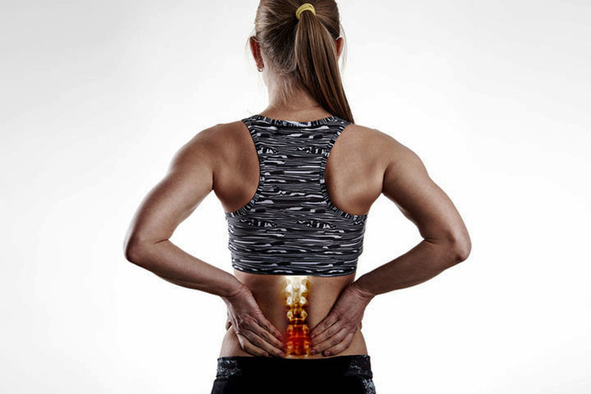
Back pain or lower back pain can manifest in two ways: as chronic back pain and can last for months or years and as acute back pain or lumbago.

The worst development of back pain is sciatica. Sciatica is pain that starts in the waist and ends in the foot at a different height each time and can reach the toes and the sole of the foot.

Sciatica is an urgent condition and should always be examined by a doctor. The risk in these cases is chronicity. If the condition remains untreated then the pain will become permanent, the loss of sensation will become complete anesthesia and the simple muscle dysfunction will develop into complete paralysis of the lower limb.

Symptoms

In terms of symptoms, acute back pain is manifested by an acute, usually sudden pain in the lower back, which reflects up to the buttocks while at the same time a reflex contraction of the lumbar muscles occurs and the patient unconsciously assumes a scoliotic (oblique) posture every time he stands up.

The pain is so strong in the beginning that it can immobilize the person for many hours in the place where it first appeared.

The cause most of the time is the lifting (lifting) of a weight in the wrong way. The person has the feeling that something has broken in his waist, he feels intense pain and gives up the weight he is holding by screaming from the pain.

Other times the seizure occurs 1 to 2 days after lifting the weight or after opening a drawer or after a long time of crouching and working at a low table or after turning the waist, etc. Usually, each patient has his own way of starting the seizure.

The crisis usually lasts for 4 to 5 days and gradually disappears.

Diagnosis

A non-urgent evaluation to control sciatica involves a physical examination and testing of the strength of the muscles, their reflexes and their senses to determine if the problem is caused by a compressed nerve. An X-ray, MRI or CT scan or electromyography (nerve conduction study) may also be needed.

Radiological examination is also necessary in cases where the symptoms persist. Because then it may not be the same known and old disease but something new such as for example a small vertebral fracture from osteoporosis that the patient has acquired but does not know. If the patient also has a fever or other general symptoms, then a hematological laboratory test will be helpful. Of course, all these examinations will be done naturally, since the patient feels a little better and can move without much pain.

Prevention

Prevention is achieved through:

  • Use a hard mattress for sleep and during sleep the posture should be on the back or side.
  • Proper postures and proper use of the body in daily activities. Those involved in sports to improve their technique.
  • Encouraging activities such as swimming, walking, cycling, etc.
  • If it is considered necessary to use special zones for intense activities, not for a long time.
  • Daily exercise with special exercises, designed by the therapist and tailored individually to each patient.
  • In case of persistent pain, consult a specialist who can identify the cause.

Treatment

From the beginning of the crisis until 15 to 20 days when he is standing, the patient should wear a belt with straps, which he can then use only when he is going to tire his waist, such as in long hours of standing or in a large travel by car.

In addition to the medications that will be used in the initial acute phase, from time to time the patient may often use an anti-inflammatory ointment which he will apply with a light massage to relax the back muscles.
Losing a few pounds, if any, helps in recovery. Kinesiotherapy and physiotherapy should be done by specialists. The best physical therapy for the spine is swimming. The more you swim in the pool or the sea, the stronger your waist and the better you feel.

Swimming will begin after the crisis is complete, in as warm a sea or pool as possible and at the beginning the patient uses a life-saving device (lifebuoy) for safety. Theoretically, if the person manages to protect his waist and does not have another crisis in the next two or three years, then he can be considered cured.

Chronic back pain usually does not end in surgery. In rare cases when it causes severe pain and is extremely persistent or when the patient has a profession in which the perfect function of his waist is necessary (such as an athlete), then he is referred to the Orthopedist for surgery.

Therapy

The therapy will depend on the final diagnosis and should be individualized to the needs of the individual.

The conservative method includes:

  • Anti-inflammatory drugs such as aspirin or ibuprofen to reduce swelling.
  • Physiotherapy, which includes exercises aimed at strengthening the muscles and restoring range of motion.
  • Physiotherapy with natural means
  • Acupuncture

Sources

Kontzaelias DA, (2011). Physiotherapy in diseases of the musculoskeletal system, Publications: University Studio Press, Thessaloniki.

σωστή θέση γραφείο

What Is The Right Position In The Office When I Work?

What Is The Right Position In The Office When I Work?

Daily questions I receive from my patients with chronic neck and waist problems are:

  1. What is the right position for me in the office when I work?
  2. Why is it important to have the right position when sitting in front of the computer?

In a developed society in which on a daily basis and for many consecutive hours we use the computer, tablets and smartphones, without the minimum training for the correct posture and ergonomics, we are called to deal with many incidents that we could have helped preventively. It is very important to know some basic things, such as what is the correct sitting position, how much you have to sit constantly and what ergonomics the space around you should have, in order to help you and not to burden your posture. Poor posture is responsible for lower back pain, neck pain, dizziness, etc.

So what you need to know when sitting in front of a computer:

  • A proper ergonomic chair can provide the right posture and support for the spine.
  • The head should be in a position where the chin is bent backwards and does not bend forward! Do not forget that the head is one of the heaviest parts of the body !!!
  • My shoulders should be pulled back and straight at the waist.
  • It is good for the elbows to be in contact with the desk and to rest on it.
  • The knees should form an angle of 90 degrees, the soles should be in full contact with the floor and should not exceed the height of the pelvis.
  • The waist, and more specifically the pelvis, should rest on the chair in such a way that there is a complete support in the lumbar spine. In case the chair does not have a support mechanism to help our waist, anatomical waist pillows with special foam material and excellent support are commercially available, with quick relief and reduction of pain from the strain on the lower back.

However, in addition to the right position that we must have when we sit in a chair, we must also take into account basic elements of proper ergonomics of the space.

  • The height of the desk should be such that the elbows form close to 90 degrees.
  • The chair should be as close as possible to the desk. This way we can easily reach all the objects on it And thus avoid minor injuries from wrong postures and strange angles that we form with our body.
  • The keyboard, mouse and anything else we use should be close to us without having to bend over to reach it again to avoid any injury.
  • The computer screen should be at the same level and slightly lower than right in front of our eyes.

If we take frequent breaks, get up and walk, we should not worry about anything. There are also specific exercises with light stretching that help during breaks. Usually every hour of office work we have to get up for at least 10 minutes to walk and do exercises. Consult your Physiotherapist, who can help you have a much more comfortable & quality life. And do not forget that “prevention is much more important than recovery”.

εργασία μυοσκελετικές παθήσεις

Work and musculoskeletal disorders. How can we deal with them?

The term musculoskeletal disorders includes health problems of the elements that contribute to movement, such as muscles, tendons, bones, cartilage, vascular system, ligaments and nerves.

Work-related musculoskeletal disorders are injuries to parts of the body such as muscles, ligaments, tendons, joints, nerves, bones and blood vessels, which are caused or exacerbated mainly by work and its effects. of the immediate work environment. Most work-related musculoskeletal disorders are cumulative diseases, due to repeated exposure to high or low-intensity stress over a long period of time. However, musculoskeletal disorders can also be acute injuries, such as fractures, that occur during an accident.

These diseases mainly affect the back, neck, shoulders and upper extremities, but can also occur in the lower extremities. Some musculoskeletal disorders, such as carpal tunnel syndrome, have clear signs and symptoms. Others present only with pain or discomfort without a clear indication of a specific condition.

Musculoskeletal disorders are the most common work-related problem in Europe. Almost 24% of workers in Europe report suffering from back and lumbar pain and 22% complain of myalgias. Both diseases are more prevalent in the new Member States, at 39% and 36% respectively.

Depending on the type of work, there are different risk factors for the body, increasing the risk of developing some musculoskeletal disorders.

RISK FACTORS

Indicative risk factors that may be responsible for the occurrence of musculoskeletal disorders are:

  • Prolonged standing
  • Prolonged sedentary work
  • Large power applications
  • Repeated movements
  • Weight lifting mistakes
  • Poor body alignment when performing the task
  • Work tool weight and tool handle
  • Work rate
  • Habits of the person (smoking, lack of exercise, etc.)
  • Somatometric characteristics of the individual (age, height, weight, etc.)
  • Psychological stress at work etc.

PREVENTION

In order to reduce the risk of disorders, first of all, on the one hand, the factors related to the employee must be improved and on the other hand, the work must be ergonomically correct and psychosocially tolerated.

Regular exercise for strengthening and flexibility of the body, adopting a healthy lifestyle, maintaining the desired body weight, creates a strong body so that it can cope with stress. On the other hand, the implementation of correct ergonomic tactics play an important role. Maintaining a generally upright posture at work, frequent breaks especially if the work is monotonous, has many repetitions, or prolonged static position, as well as the correct lifting of loads are some of the basic things that need to be taken care of. Also, the lack of pressure in the workplace and good cooperation with colleagues is essential.

Unfortunately, despite the advancement of science and the possibilities for safe and productive work at work, work-related musculoskeletal disorders are a major problem worldwide, constantly worsening even in the most economically developed countries. The modern way of life, smoking, poor diet, stress and lack of exercise as well as working conditions that are mostly characterized by intensification, flexible hours, overtime and measures such as the abolition of the Sunday holiday and raising age limits retirement contributes to the exacerbation and perpetuation of the phenomenon if we do not take action.

The solution to deal with this phenomenon is in the hands of every human being. In order to achieve prevention and to have a drastic reduction of work-related musculoskeletal diseases, it is crucial that workers collectively work with health professionals to improve their working conditions and to create free public sports and leisure facilities. contribute to improving health and quality of life.

REHABILITATION

The rehabilitation of such diseases includes the treatment of the symptoms and the restoration of strength and neuromuscular coordination with exercises and natural means in order to restore the functionality of the affected area. In addition, proper ergonomics and body function are learned for daily life activities to reduce the risk of re-injury.

Essentially, the treatment of musculoskeletal disorders requires an integrated management approach. This approach should include not only the prevention of new cases, but also the stay at work, the rehabilitation and reintegration of workers already suffering from musculoskeletal disorders.

Sources

Kontzaelias DA, (2011). Physiotherapy in diseases of the musculoskeletal system, Publications: UniversityStudioPress, Thessaloniki.