Address
1. 70 Asklipiou Street, Mytilene, 81100
2. 7 Myraktis Street, Nea Smyrni, 17121
Working Hours
Mon-Fri: 09:00-21:00
Saturday:10:00-14:00

Lorem ipsum dolor sit amet, consectetur adipiscing elit. Nam bibendum ultrices arcu ac vestibulum. Nam nec tempus quam.

[fusion_separator style_type=”none” sep_color=”” border_size=”0″ icon=”” icon_circle=”” icon_circle_color=”” alignment=”center” hide_on_mobile=”small-visibility,medium-visibility,large-visibility” class=”” id=”” top_margin=”10px” bottom_margin=”10px” /]

[fusion_button link=”https://physiogalinos.gr/our-history/” text_transform=”” title=”” target=”_self” link_attributes=”” alignment=”left” modal=”” color=”default” button_gradient_top_color=”” button_gradient_bottom_color=”” button_gradient_top_color_hover=”” button_gradient_bottom_color_hover=”” accent_color=”” accent_hover_color=”” type=”” bevel_color=”” border_width=”” size=”” stretch=”default” shape=”” icon=”” icon_position=”left” icon_divider=”no” animation_type=”” animation_direction=”left” animation_speed=”0.3″ animation_offset=”” hide_on_mobile=”small-visibility,medium-visibility,large-visibility” class=”” id=””]Our History[/fusion_button]

ρήξη μηνίσκου

Meniscus Tear

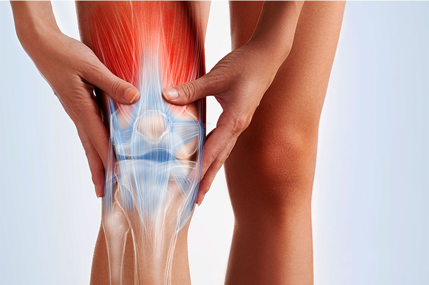
Meniscus tear is one of the most common causes of knee pain. The knee joint consists of the peripheral end of the femur, the central end of the tibia and the patella. Each knee has 2 menisci, the inner meniscus on the inside of the knee and the outer meniscus on the outside of the knee.

The menisci are elastic fibrochondrial formations that consist of water and collagen. They have a crescent shape and act as a “shock absorber” absorbing vibrations during the movement of the knee and distributing the weight evenly throughout the joint. The menisci also contribute to the stability and lubrication of the knee.

How does meniscus tear occur?

Meniscus tear is usually traumatic. The force required to tear depends on the size of the injury and the pre-existing meniscus degeneration. It usually occurs in young patients during a sports activity, but it can also occur in any other activity when they exert rotational and compressive loads that exceed the strength of the meniscus. Older patients with pre-existing degenerative lesions are also vulnerable and are more likely to have a meniscus tear even during daily movement.

Symptoms of meniscus tear

At the time of injury, the patient may feel or even hear a noise as if something had broken. Sometimes it is possible for the patient to continue his activity but gradually the knee may swell and become stiff.

The most common symptoms are:

  • Pain in the inside or outside of the knee (corresponding to a broken meniscus) that worsens with the knee bent (as in the deep seat or stairs)
  • Swelling and stiffness
  • Knee bruise or involvement when there is a dislocated piece
  • Feeling of instability

Diagnosis

The diagnosis is made by history and clinical examination with special techniques. MRI serves to confirm the diagnosis, to rule out other lesions and to plan treatment.

Treatment

In case the symptoms are mild, conservative treatment is followed, which includes medication and physiotherapy to prevent stiffness, reduce swelling and avoid muscle atrophy. The patient must follow a special program of strengthening the muscles that contribute to the stability of the joint.

Physiotherapy includes:

  • Electrotherapy
  • Ultrasound
  • Ice therapy
  • Manual therapy
  • Acupuncture
  • Diamagnetic pump
  • BFR
  • Predisposition and balance exercises

Immediate treatment of the problem can significantly reduce the chances of surgery, unless the extent of the injury is large. In case the conservative treatment fails, the patient should resort to arthroscopic treatment.
ρήξη αχιλλείου τένοντα 2

Achilles Tendon Rupture

What exactly is a partial rupture of the Achilles tendon?

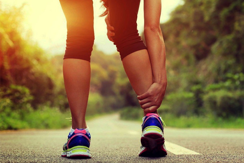
The group of muscles in the back of our lower leg is called the calf. The calf consists of two main muscles, one of which protrudes above the knee joint (gastrocnemius) and the other below the knee (tibia). Both of these muscles are located in the heel bone through the Achilles tendon.

When the calf muscles contract, force is also exerted on the Achilles tendon. When the contraction is intense due to multiple repetitions or intense effort, it can cause the Achilles tendon to rupture.

ρήξη αχίλλειου τένοντα 1

Ruptures in the Achilles tendon range from a small, partial rupture with minimal pain and difficulty moving to a total rupture, which may require surgical repair.

Cause

Achilles fractures usually occur when the patient tries to accelerate from a standstill or when he rushes sharply forward, such as during a game of tennis, football, volleyball, etc.

Symptoms

Patients with partial rupture usually experience pain during activities such as walking (especially uphill), climbing stairs, running and jumping. It is also common for them to feel pain at rest after such activities, especially when they wake up in the morning. Swelling or tenderness may occur.

Diagnosis

The diagnosis is usually made during a clinical examination and confirmed by magnetic resonance imaging or ultrasound.

Treatment

Most patients with an Achilles tendon rupture are treated with a physical therapy program. The success rate of this program depends on the patient’s compliance with the program. One of the main keys of the program is to stop the patient any activity that increases the pain, until the symptoms completely subside. This allows the body to begin the healing process, as there will be no further tissue damage. Once this is achieved, a gradual return to these activities is advisable, as long as there is no increase in symptoms.

In the initial phase the inflammation can subside with anti-inflammatory and regular ice packs. To ensure the best possible result it is very important a program of restoration of flexibility and strengthening, under the supervision of a physiotherapist.

Factors Contributing to Achilles Tendon Rupture

There are several factors that can predispose to rupture of the Achilles tendon. These need to be assessed and corrected under the guidance of a physiotherapist. Some of the factors that contribute to this condition are: lack of flexibility, wrong way of exercising, insufficient mechanical support, posture, insufficient warm-up and muscle weakness.

Physiotherapy for Partial Rupture of the Achilles Tendon

Physiotherapy for Achilles tendon rupture is valuable for speeding up recovery, ensuring a very good result and reducing the chance of re-injury. Physiotherapy includes:

  • Soft molecule techniques (therapeutic massage)
  • Electrotherapy
  • Taping
  • Exercises to strengthen and improve muscle elasticity
  • Retraining
  • Articular Mobilization (manual therapy)
λουτροθεραπεία μυτιλήνη

Thermal Spa Therapy – Medical Observations and Applications. Benefits of Thermalism and Hydrotherapy.

By thermal bath therapy we mean the application of thermal waters in the healing of human. This type of treatment is part of physiotherapy, ie the use of natural remedies in therapy.

Hydrotherapy includes the following forms: bath therapy, positherapy, inhalation therapy, sludge therapy or mud therapy (mud baths), as well as thalassotherapy.

Spa therapy is based on the following main properties of the thermal water: the thermal stimulus, the mechanical stimulus, and finally, the effect of the physicochemical components of the thermal water. This type of treatment is part of physiotherapy, ie the use of natural remedies in therapy.

Medical Observations

There are general rules that follow spa therapy, based on general medical observations.

More detail:

  • Before the start of the treatment, the coexistence of other diseases that exclude or limit the thermal bath therapy in terms of duration, temperature, methods of application, etc. will be studied.
  • Rheumatic disease will be studied and the duration, temperature, type of treatment will be determined accordingly (ie bath, hydromassage, steam baths, mud baths, hydrokinesiotherapy), as well as their area of ​​application (such as neck, waist, shoulder, arms, hip, knee, etc.).
  • It is examined if the patient is receiving any treatment that usually should not be stopped.
  • Finally, a review is performed in the middle of the three-week treatment and at the end. The patient is provided with a letter from the bath doctor with his remarks about the treating doctor.
  • The various treatments are usually done in the morning and last from a few minutes to an hour depending on the type.

Applications

The ways of using the thermal water are based on the action factors of the thermal water. That is:

By inhalation, radon penetrates the body.

In positherapy with hypotonic (oligometallic) and isotonic waters, diuretic, cholagogue and metabolism in general appear. Positherapy is also of interest to many rheumatic diseases, such as gout and possibly degenerative arthropathy. The waters of Loutraki, Kaiafa, Platistomou, Nigrita, Sariza, Souroti, Xinou Nero, etc. are used.

With external techniques, which find great application in rheumatic diseases.

More specifically:

  • The bath is full body, semi-bath, local, at temperatures 34 – 36 ° C. It is performed in an individual bath, ordinary or spacious, but also in a swimming pool. It can be combined with sprinkling or hydromassage, which stimulate the muscles.
  • The steam bath that can be local or universal.
  • The mud bath that is also all over or more usually local (poultice) in the affected area (joints). The action of the mud is mainly attributed to the high temperatures of 40 – 46οC and more. Heat inducers are known, that solid bodies retain heat for a long time and that then the human body tolerates it more easily.
  • This is why mud is used for action on deep organs (hip, etc.). But thermal mud is not a simple mixture of solid substrate and thermal water. The soil is selected from volcanic soils, from alluviums and swamps. It is cultivated with thermal water for many months or years and undergoes fermentation treatments. Thus, it acquires physicochemical properties in the presence of living organisms that facilitate the absorption of sulfur and radon, inflammatory elements and the reduction of pain.
  • A remarkable way of applying the thermal water is its use in swimming pools, which is the currently accepted by everyone hydrokinesiotherapy. This method exploits all three factors of thermal water – chemical, thermal and mechanical. Hydrokinetic therapy is highly valued for the prevention of permanent damage (such as pathological lesions, atrophies, deformities, ankylosis) but also for their restoration as much as possible.

Benefits

The beneficial effect usually comes a few weeks after the end of the thermal bath therapy but often during it. Some recommend partial spa therapy at the beginning and end of the bath period.

Bath Reaction

Complementary physiotherapy may sometimes be needed, such as mechanotherapy or laboratory examination of the patient. The person dealing with the object must be aware of the possible bath reaction. This may occur during the first 5 to 10 days of spa treatment and consists of a few days and a temporary clinical deterioration of the patient’s general condition, possibly with fever, anorexia, headaches and insomnia. The bath reaction may be local with worsening of local symptoms and has nothing to do with the final outcome of treatment. Especially in cases of gout can cause a new crisis despite taking colchicine. The patient who is going to undergo thermal bath therapy must be informed about these phenomena.

SOUSCES

Angelidis Z., (2007). Thermal natural resources and thermalism, Eleftherios Kordelios Environmental Education Center, Thessaloniki.

Boleti P., (2006). Thermal Tourism: The Greek perspective with the emphasis on education and training. International Conference of Trends, Impacts and Policies on Tourism, Edipsos.

Gerbod P., (2004). Loisirs and santé: the thermalisms in Europe of the origins of our days, Paris: Honoré Champion.

Hare J.P., Corrall R.G.M., Dieppe P.A., Evans J.M., Heywood A., Lunn G., Summerhayes C., & Walters G., (1985). Observations on the effects of immersion in Bath spa water, British Med., J. 291: 1747 – 1751.

Muscle Strain

Strain is a muscle injury. The most common strains are strains of the muscles of the lower extremities, such as the strain of the thigh muscles, and the strain of the gastrocnemius (calf). There is no difference in gender and age.

Strains are divided into three categories:

First degree strains: small rupture of muscle elements.

Second degree strains: partial rupture of muscle elements, but maintenance of muscle continuity.

Third degree strains: complete cross section of the muscle, ie the muscle is cut.

θλάση κατηγορίες

Cause of a muscle strain

Muscles can be injured in two ways, by direct blows, or by over-stretching. The result is a rupture of muscle fibers (which can reach a full cross section of the muscle) and the formation of a hematoma.

The most common cause of a strain is a local injury to the muscle area, or to the myotendinous area near the joints. People who engage in sports activities are more likely to have strains. Also, people who have had strains in the past are more likely to have strains again in the same area.

There are several factors that contribute to muscle spasm:

  • Poorly prepared muscles due to poor training or poor warm-up during exercise.
  • Weak muscle due to previous injury and poor recovery.
  • The muscle has developed scar tissue inelastic at the site of the previous strain.
  • The muscle is overloaded and has reached a point of great fatigue.
  • Tight muscles or muscles exposed to cold are more easily injured.

Preventing during sports activities and strengthening the areas that have a history of strains with elastic bandages, reduces the chances of muscle injury.

Symptoms

Obviously, the symptoms depend on the severity of the injury. In the case of muscle strain, the rupture of the muscle fibers is accompanied by the leakage of blood, which accumulates in the tissues causing hematoma, the dimensions of which are proportional to the severity of the injury. The main symptom is pain at rest, which is aggravated by the effort to move the muscle concerned and is accompanied by functional disability, making it impossible to use the affected muscle.

Muscle strains occur more in the area where the muscle connects to the tendon and less in the main muscle mass. The main symptoms in these conditions are immediate pain, local tenderness in the area, the development of edema (swelling), as well as the restriction of movement in the nearby joints. Ecchymosis (bruising) can often be seen immediately in the area of ​​the strain. This is also called a hematoma.

Diagnosis

Clinical examination is the main diagnostic tool in muscle strains. The use of ultrasound and magnetic resonance imaging in recent years complements the clinical examination and mainly helps to determine the size of the injury.

Complications

Complications after a muscle strain usually occur in cases of inadequate or incomplete treatment. They concern the increasing formation of scar tissue in the area of ​​the strain, the creation of an encapsulated hematoma and the creation of bone tissue (ossifying myositis) which often leads to the limited functionality of the muscle and the joint in which it affects. Surgical treatment is usually the method of choice in these cases.

Treatment of a muscle strain

Conservative treatment is first and foremost the first approach to treating strains.

It consists of:

  • Bandaging the muscle (upper or lower limb) with an elastic bandage
  • Use of rest splints
  • Administration of non-steroidal anti-inflammatory drugs
  • Laying ice for the first two days
  • Physiotherapy for rehabilitation, if necessary
  • Second and third degree strains are assessed by the doctor and depending on other possible lesions, surgery (muscle suturing)

The physiotherapist with the means at his disposal, for the first 24 hours tries to reduce the swelling and pain using TECAR, TENS currents, cross currents and cryotherapy.

After the first 24 hours, the physiotherapist’s goal is to increase blood flow to the site of injury (via TECAR, ultrasound, hot pads, diathermy and laser) to achieve the maximum degree of nutrient supply for wound healing.

Then a program of stretching exercises is imposed on the injured muscle. After the rehabilitation, the isotonic exercises for additional strengthening gradually begin.

Following are specialized exercises of susceptibility and neuromuscular fitting, ie exercises of retraining the muscles for a safe return to daily activity.

SOURCES
Evans W.J., & Cannon J.G., (1991). The metabolic effects of exercise in muscle damage, Exercise Sport Sc. Rev, (19): 99 – 125.

παγωμένος ώμος

Frozen Shoulder

What is a frozen shoulder?

Frozen shoulder is a condition characterized by inflammation and stiffness of the connective tissue that covers the shoulder joint.

Sometimes the articular pock of the shoulder joint becomes inflamed and “tightens” the joint. When this happens then we have frozen shoulder syndrome.

frozen shoulder

In general, frozen shoulder syndrome can be divided into three stages:

  • Painful stage: In the first stage there is pain in all movements of the shoulder. Also at this stage the shoulder stiffness begins.
  • Stiffness stage: The second stage of the frozen shoulder is characterized by a great lack of movement, due to the scar tissue that develops in the joint pocket. Patients have great difficulty reaching the hand back to their back. The pain at this stage begins and subsides.
  • Stage of remission of symptoms: At this stage the symptoms begin to subside as the shoulder becomes more relaxed and moves comfortably.

Causes

Although the exact cause is not known, some injury to the shoulder joint or adjacent tissues is thought to be responsible. Frozen shoulder is more common in cases where proper treatment has not been applied. It is also more common in cases where the shoulder has been immobilized for a long time (eg after surgery), if the patient has diabetes or another autoimmune disease.

Symptoms

Symptoms associated with a frozen shoulder develop gradually. Initially, patients experience a shoulder discomfort that develops into “sharp” pain with specific movements or activities. The pain is deep in the shoulder, although sometimes there is discomfort in both the arm and the neck. In the above areas there is stiffness. The pain associated with frozen shoulder syndrome increases with all shoulder movements.

Activities such as raising the hand, carrying, lifting objects, pushing or pulling, shoulder pressure during sleep, loosening a bra increase the symptoms. A common phenomenon is night pain and pain in the first days of the day. When we move to the second stage of the frozen shoulder then the pain begins to decrease. In addition to pain, patients with frozen shoulder have a high degree of stiffness in the shoulder joint. Stiffness affects all shoulder movements but more the lifting and turning.

Diagnosis

Subjective (history taking) and objective (clinical examination) evaluation are sufficient to lead the physiotherapist to diagnose frozen shoulder syndrome. Sometimes tests such as MRI scans can help.

Treatment

Once frozen shoulder syndrome appears, a few things can be done to speed up recovery. The best treatment is prevention. For this reason, any shoulder injury must be completely healed, according to the treatment protocols prepared by the physiotherapist in each case.

The goal of treatment once frozen syndrome is established is to maintain shoulder mobility and strength, as well as reduce pain. It is very important to avoid activities and increase the symptoms. This helps our body heal damaged tissues and reduce inflammation.

Prognosis

In some cases the frozen shoulder subsides after months. Rarely do the symptoms go away after eighteen months or more. The painful stage usually lasts from two to six months. The stage of stiffness lasts four to twelve months and the last stage of recovery four to eighteen months.

Prognostic factors of frozen shoulder syndrome

There are several factors that contribute to the development of frozen shoulder. They all need to be evaluated and corrected during rehabilitation by the physiotherapist. Some of them are:

  • History of shoulder surgery
  • Inadequate / inadequate treatment after injury or shoulder surgery
  • Diabetes
  • Autoimmune Diseases
  • Age over 40

Physiotherapy for frozen shoulder syndrome

  • Soft molecule techniques (massage)
  • Electrotherapy
  • Taping
  • Exercises to improve the elasticity of the shoulder muscles
  • Education – tips
  • Articular mobilization (manual therapy)
  • Hydrotherapy
  • Dry needle technique
  • Thermotherapy